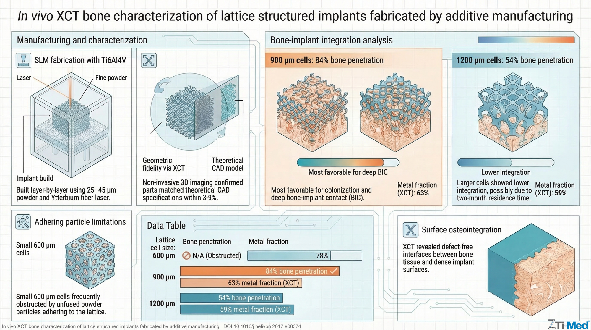
In vivo XCT bone characterization of lattice structured implants fabricated by additive manufacturing
Abstract
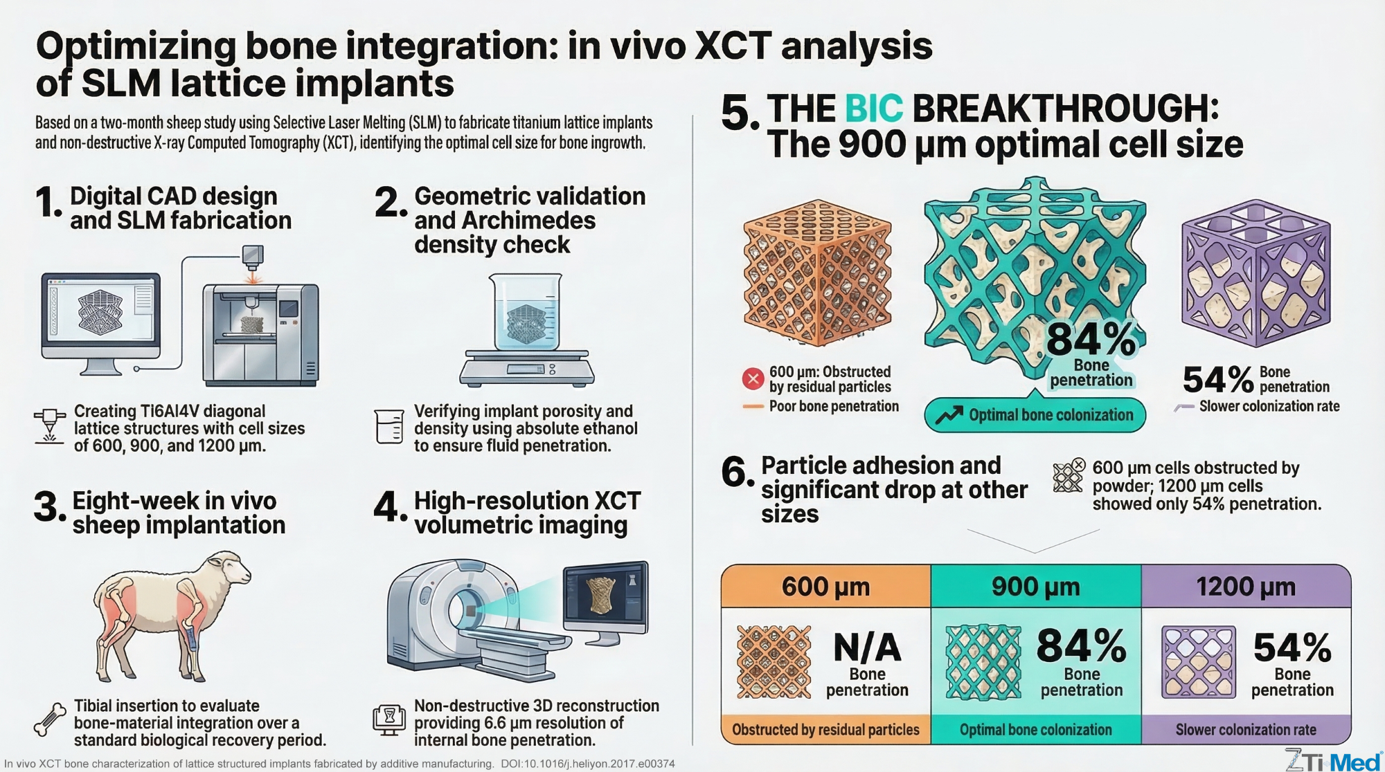
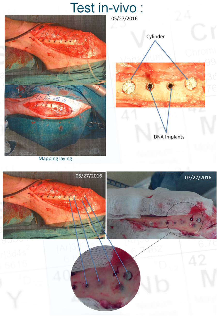
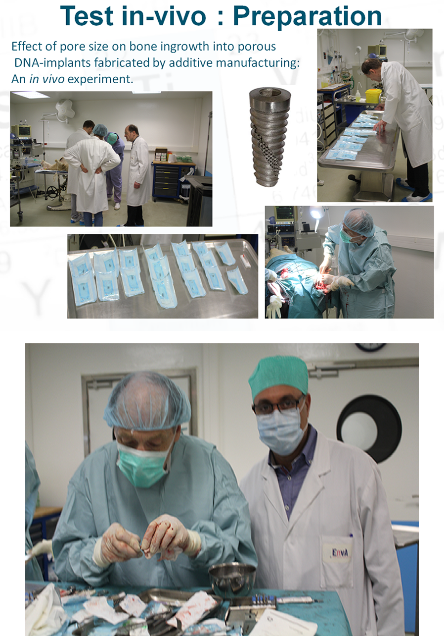
Several cylindrical specimens and dental implants, presenting diagonal lattice structures with different cell sizes (600, 900 and 1200 μm) were additively manufactured by selective laser melting process. Then they were implanted for two months in a sheep. After removal, they were studied by Archimedes’ method as well as X-ray computed tomography in order to assess the penetration of bone into the lattice. We observed that the additive manufactured parts were geometrically conformed to the theoretical specifications. However, several particles were left adhering to the surface of the lattice, thereby partly or entirely obstructing the cells. Nevertheless, bone penetration was clearly visible. We conclude that the 900 μm lattice cell size is more favourable to bone penetration than the 1200 μm lattice cell size, as the bone penetration is 84% for 900 μm against 54% for 1200 μm cell structures. The lower bone penetration value for the 1200 μm lattice cell could possibly be attributed to the short residence time in the sheep. Our results lead to the conclusion that lattice implants additively manufactured by selective laser melting enable better bone integration.
In vivo XCT bone characterization of lattice structured implants fabricated by additive manufacturing
Abstract
Several cylindrical specimens and dental implants, presenting diagonal lattice structures with different cell sizes (600, 900 and 1200 μm) were additively manufactured by selective laser melting process. Then they were implanted for two months in a sheep. After removal, they were studied by Archimedes’ method as well as X-ray computed tomography in order to assess the penetration of bone into the lattice. We observed that the additive manufactured parts were geometrically conformed to the theoretical specifications. However, several particles were left adhering to the surface of the lattice, thereby partly or entirely obstructing the cells. Nevertheless, bone penetration was clearly visible. We conclude that the 900 μm lattice cell size is more favourable to bone penetration than the 1200 μm lattice cell size, as the bone penetration is 84% for 900 μm against 54% for 1200 μm cell structures. The lower bone penetration value for the 1200 μm lattice cell could possibly be attributed to the short residence time in the sheep. Our results lead to the conclusion that lattice implants additively manufactured by selective laser melting enable better bone integration.
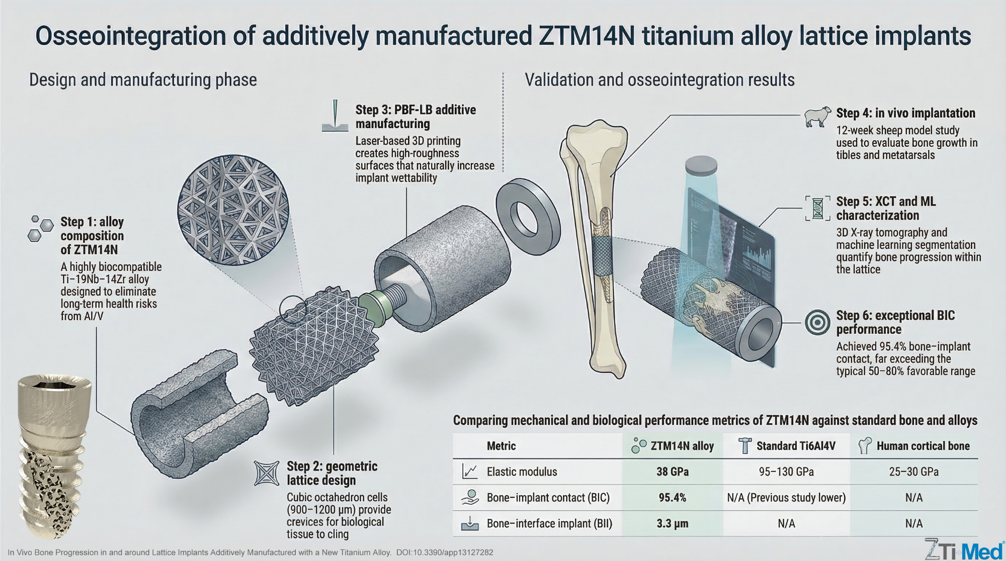
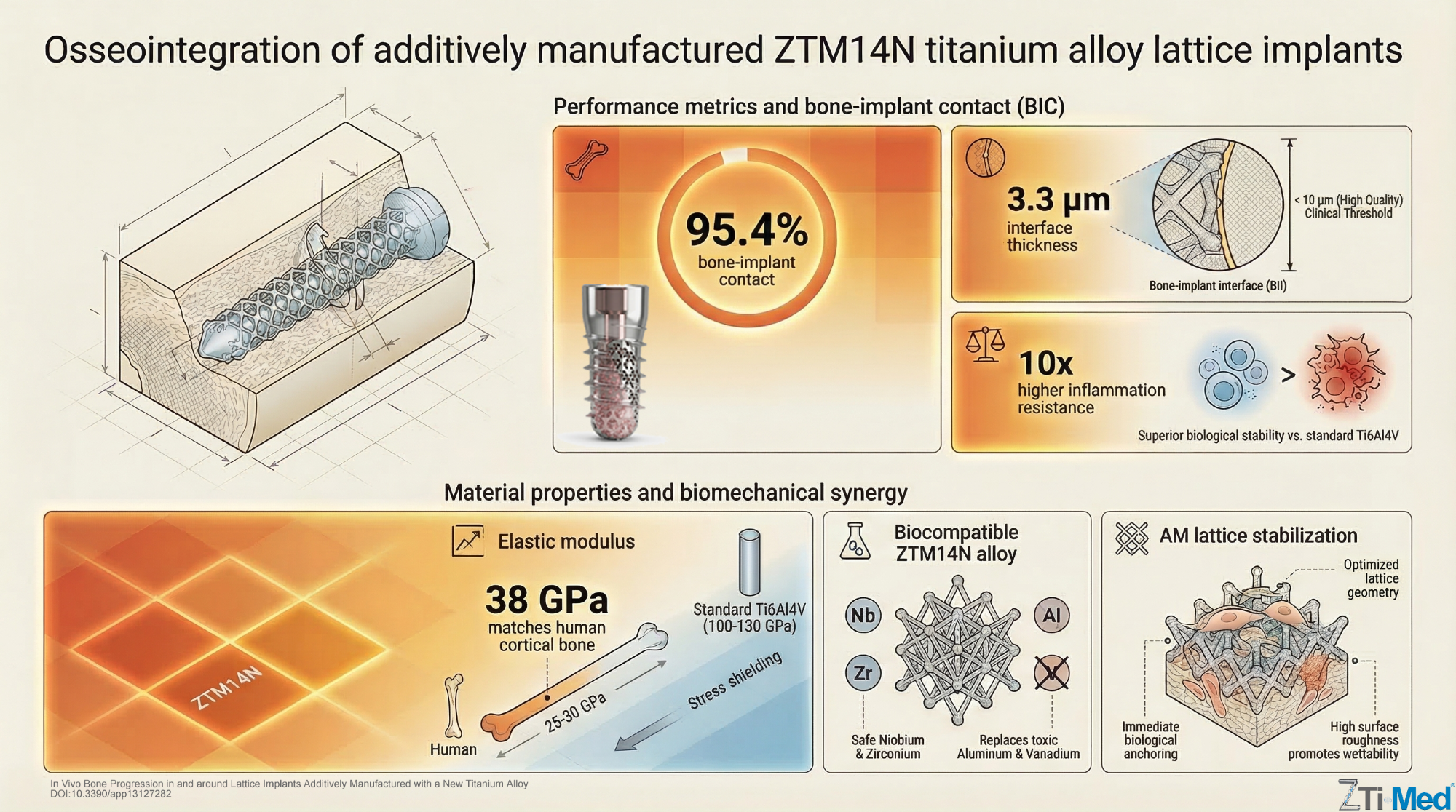
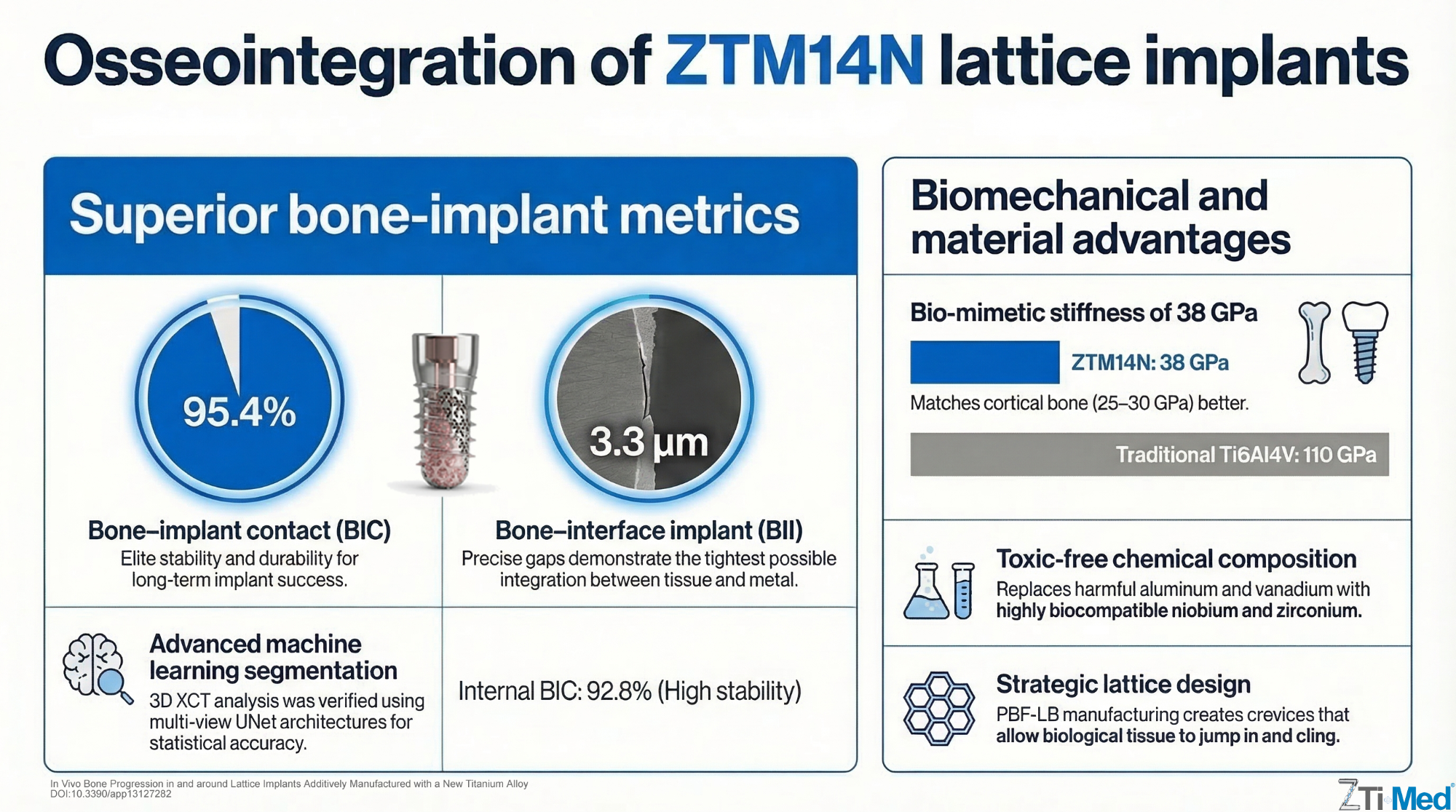
In vivo bone progression in and around lattice implants additively manufactured with a new titanium alloy
Abstract
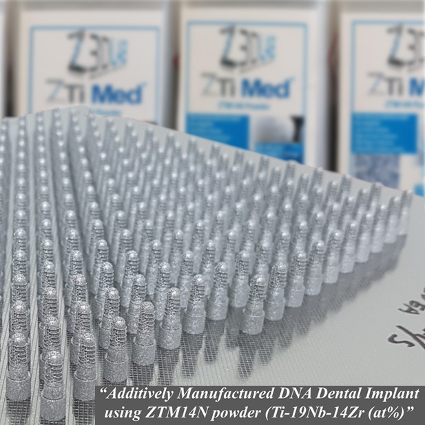
The osseointegration process in and around additively manufactured (AM) lattice structures of a new titanium alloy, Ti–19Nb–14Zr, was evaluated. Three different implants, including lattices with increasing high sidewalls gradually closing them, were designed, manufactured and implanted in the tibia and metatarsal bone of two sheep for twelve weeks. After removal, they were characterized with X-ray computed tomography (XCT). The 3D XCT images were segmented using machine learning. The bone-interface implant (BII) and bone-implant contact (BIC) were studied. The results show that, since AM naturally leads to high roughness surface finish, the wettability of the implant is increased. The new alloy possesses an increased affinity to the bone enhancing the quality of osseointegration. The lattice provides crevices, in which the biological tissue can jump in and cling. The combination of these factors is pushing ossification beyond its natural limits. Therefore, the quality and speed of the ossification and osseointegration in and around these Ti–19Nb–14Zr AM laterally closed lattice implants open the possibility of bone spline key of prostheses. This enables the stabilization of the implant into the bone while keeping the possibility of punctual hooks allowing the implant to be removed more easily if required.
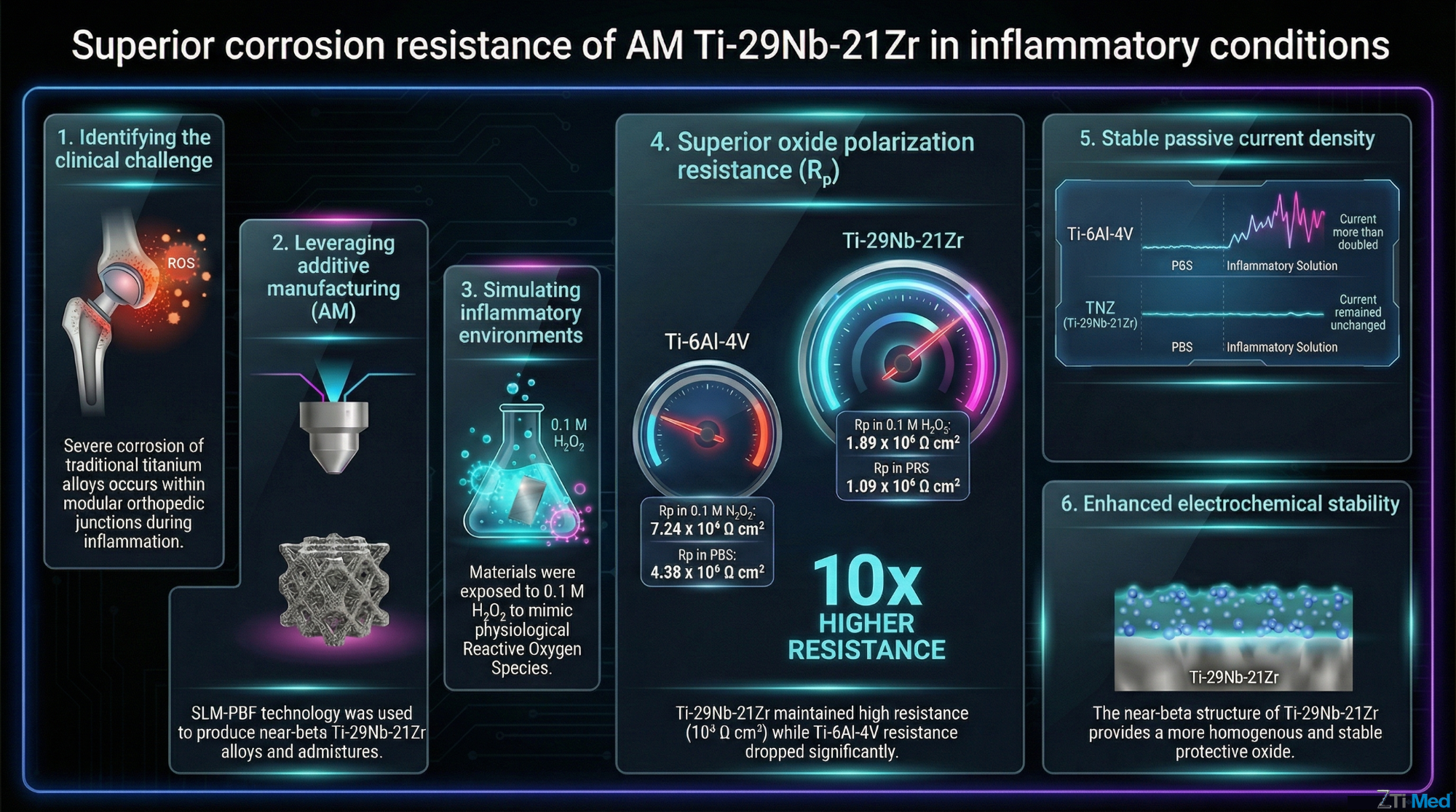
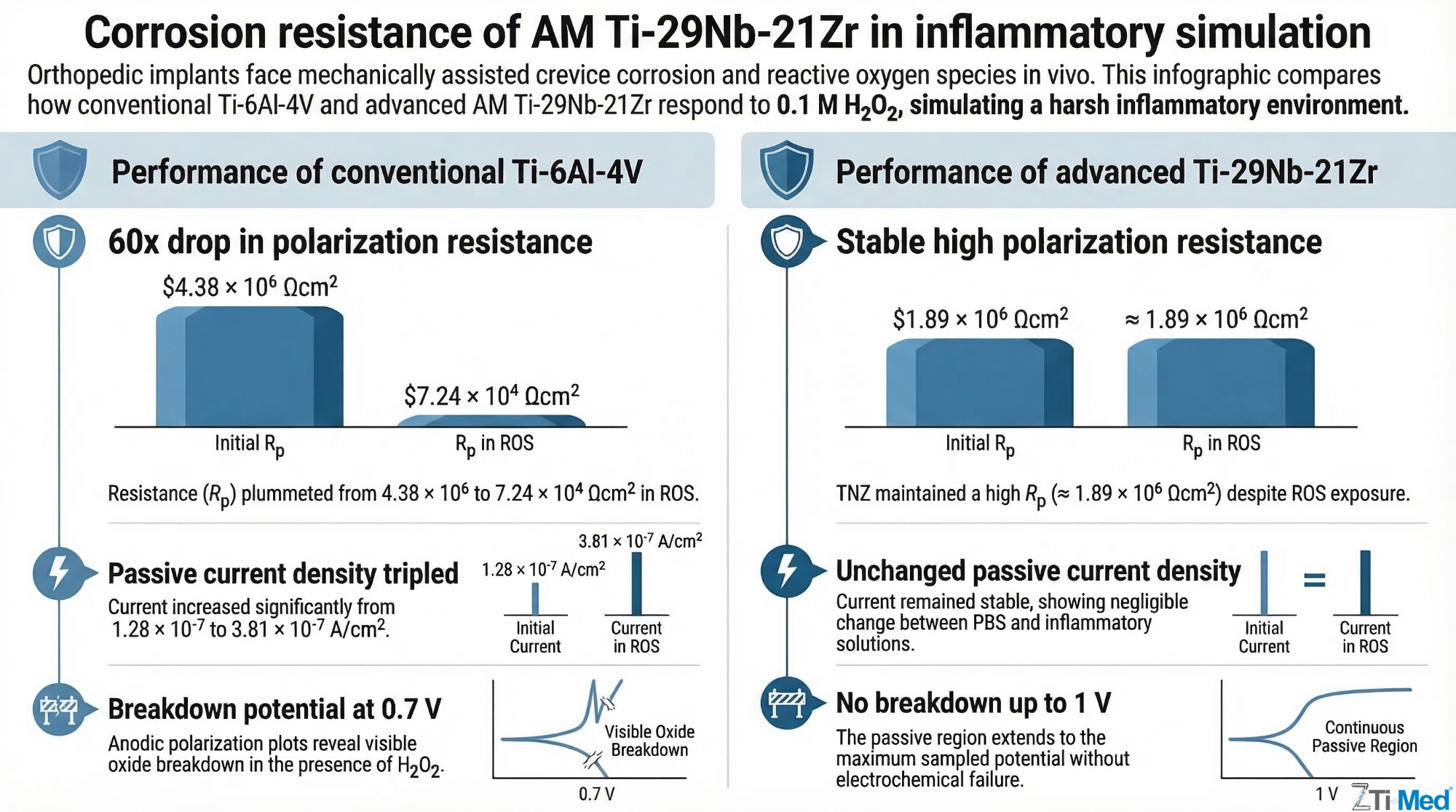
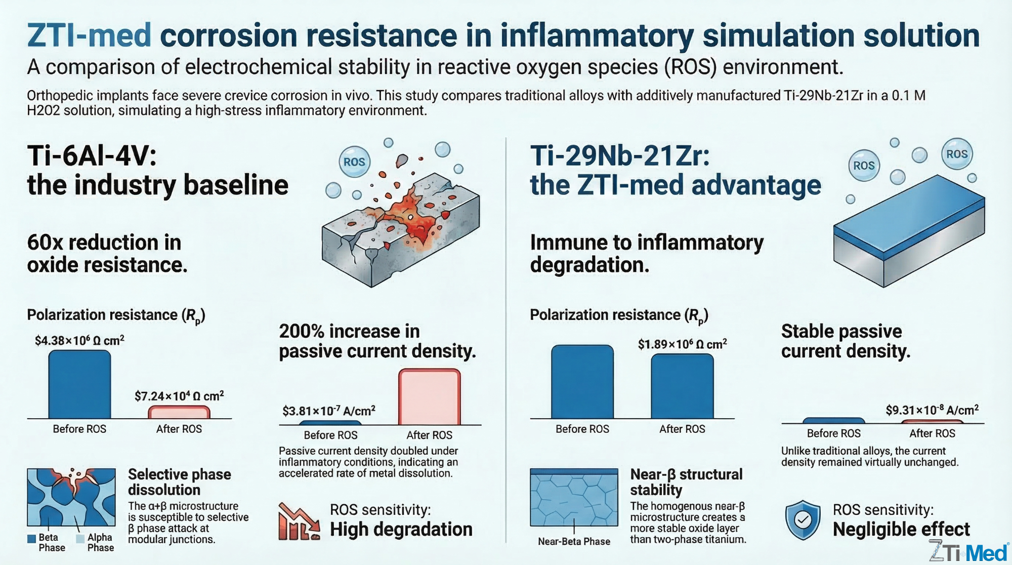
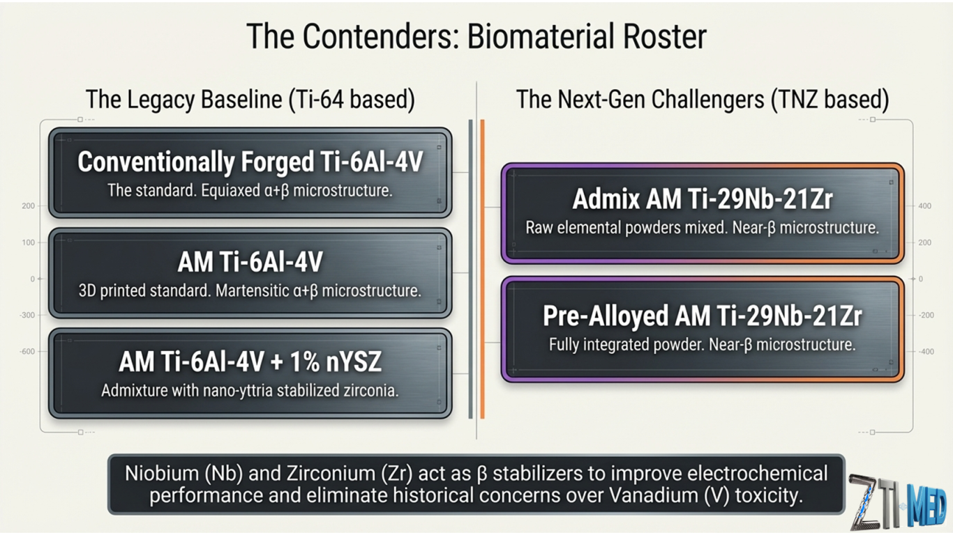
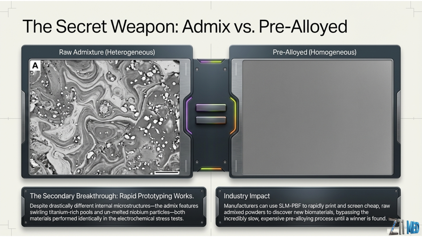
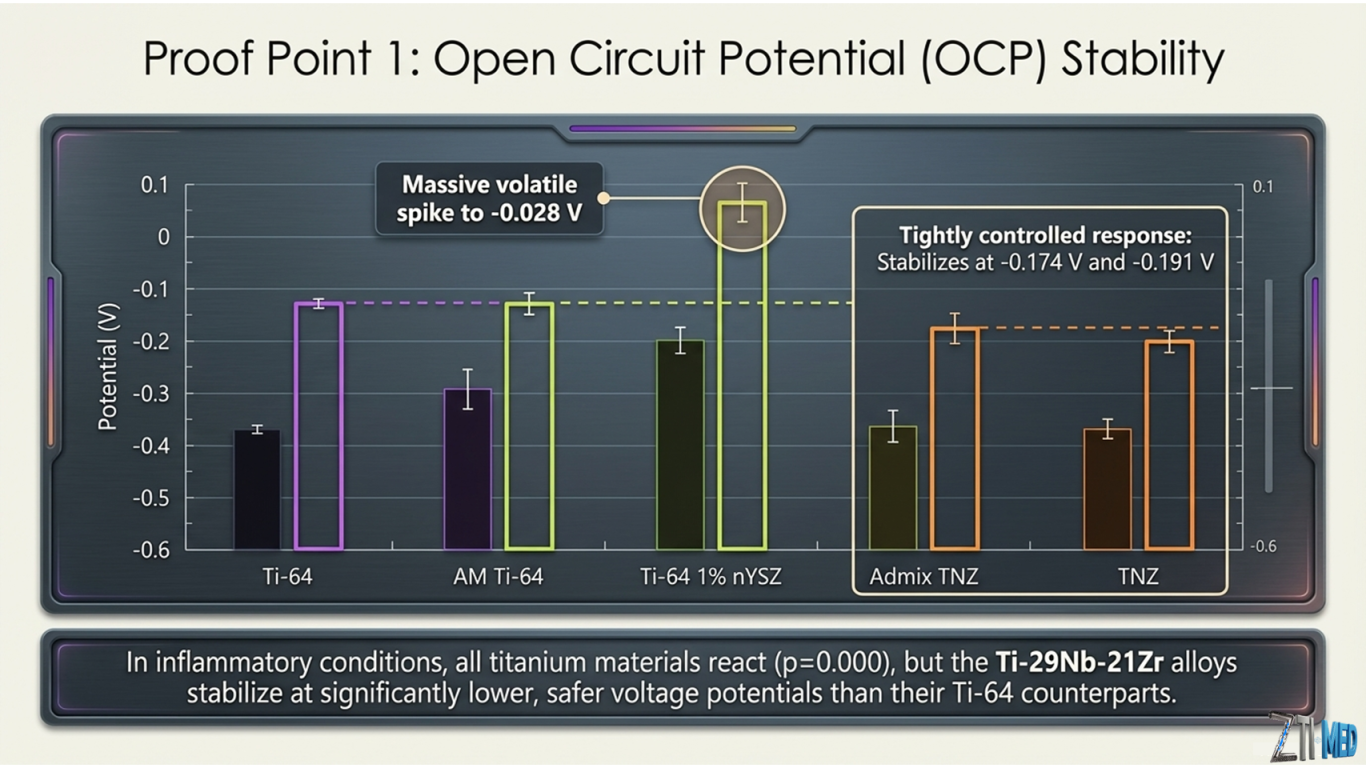
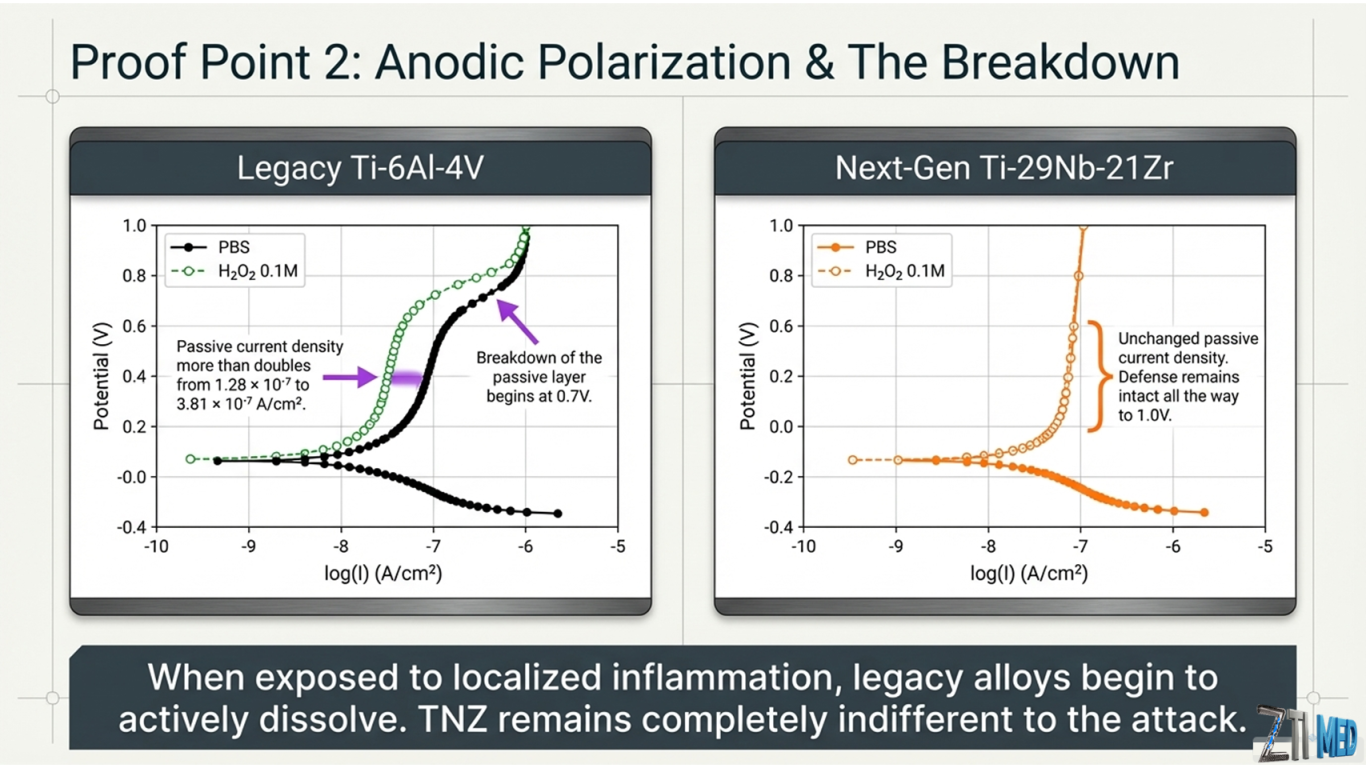
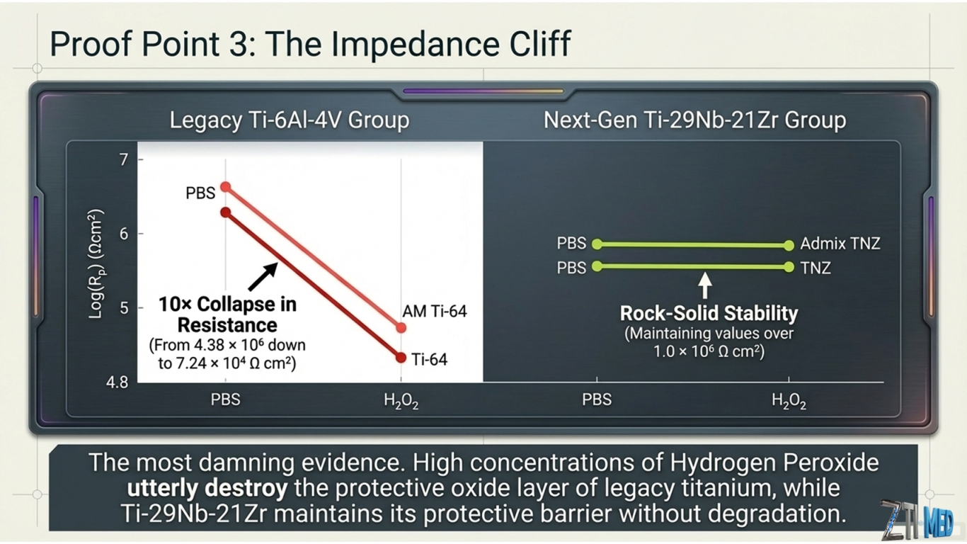
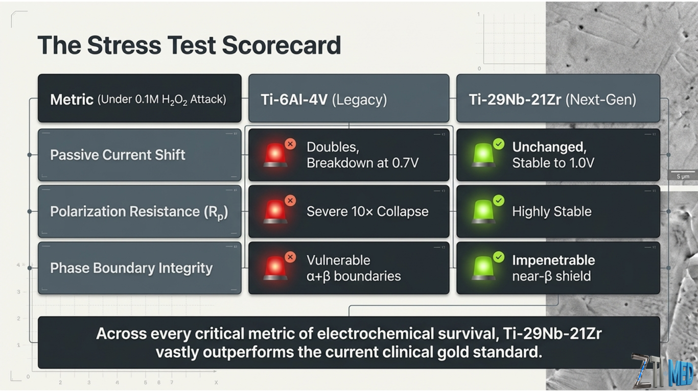
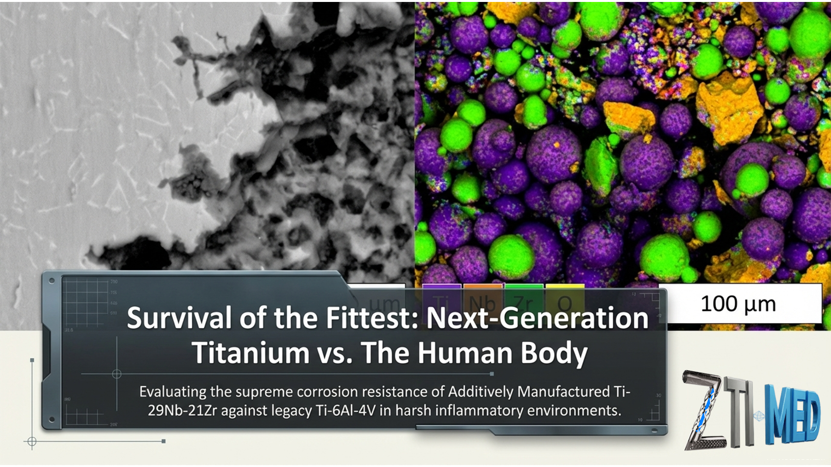
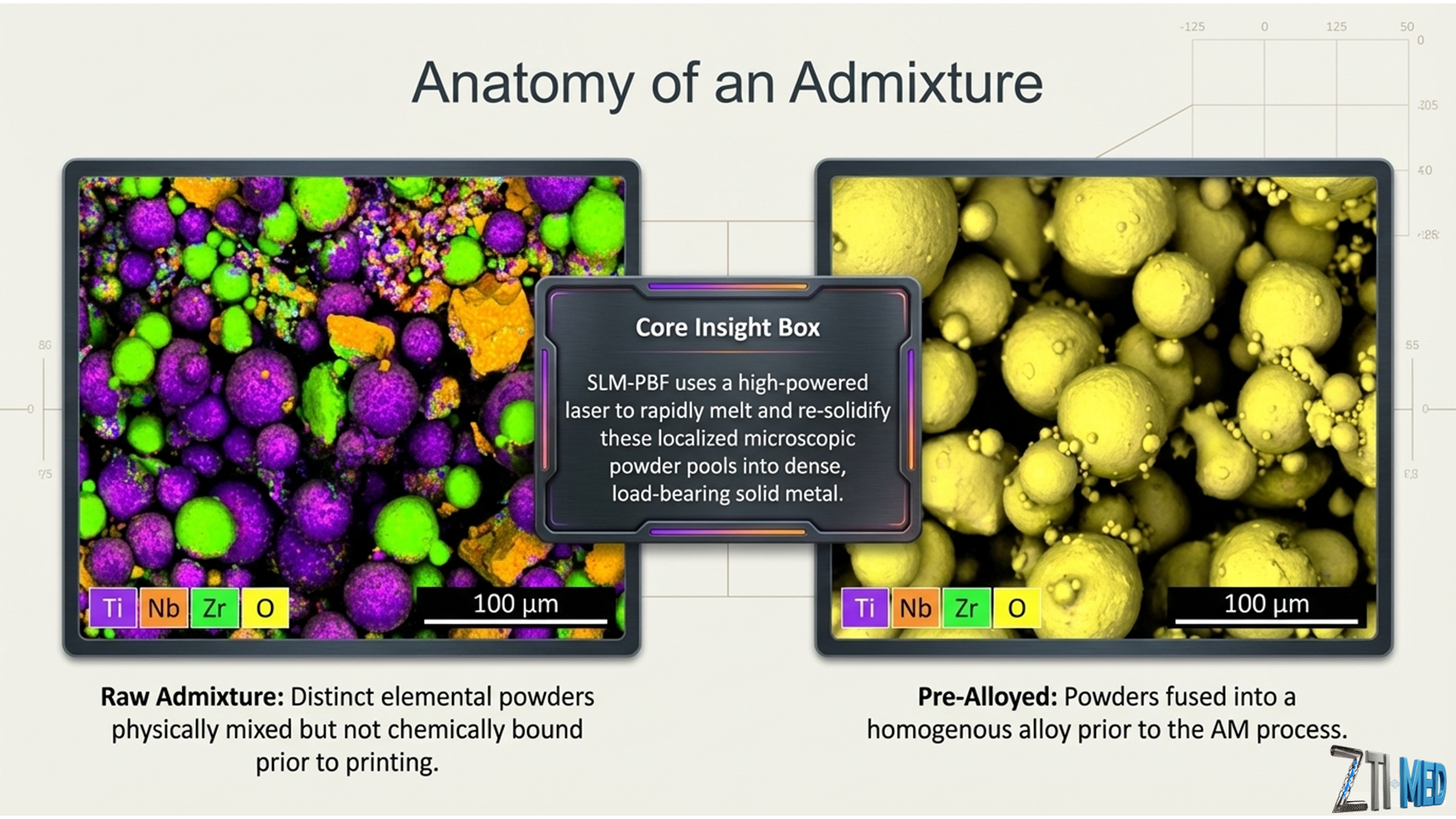
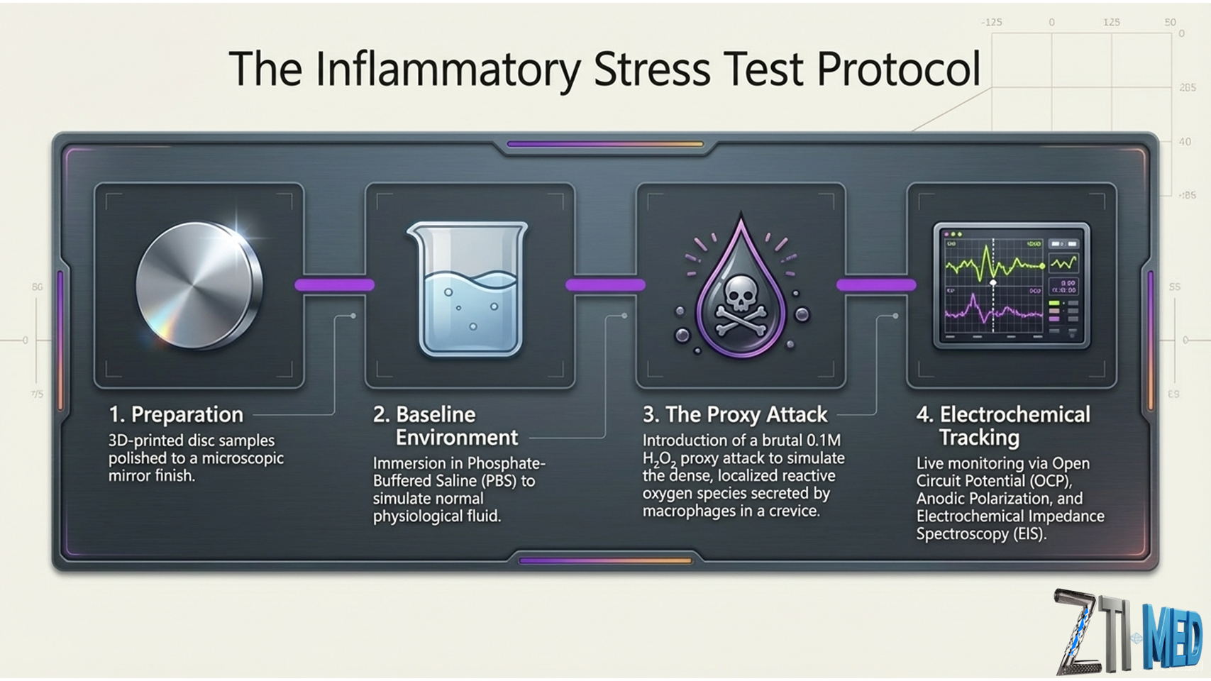
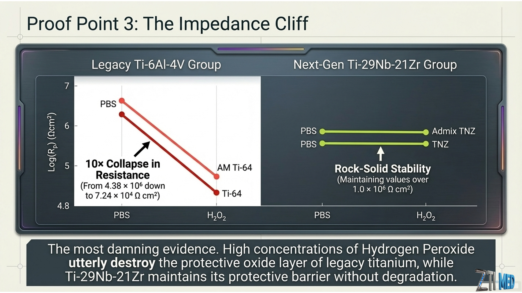
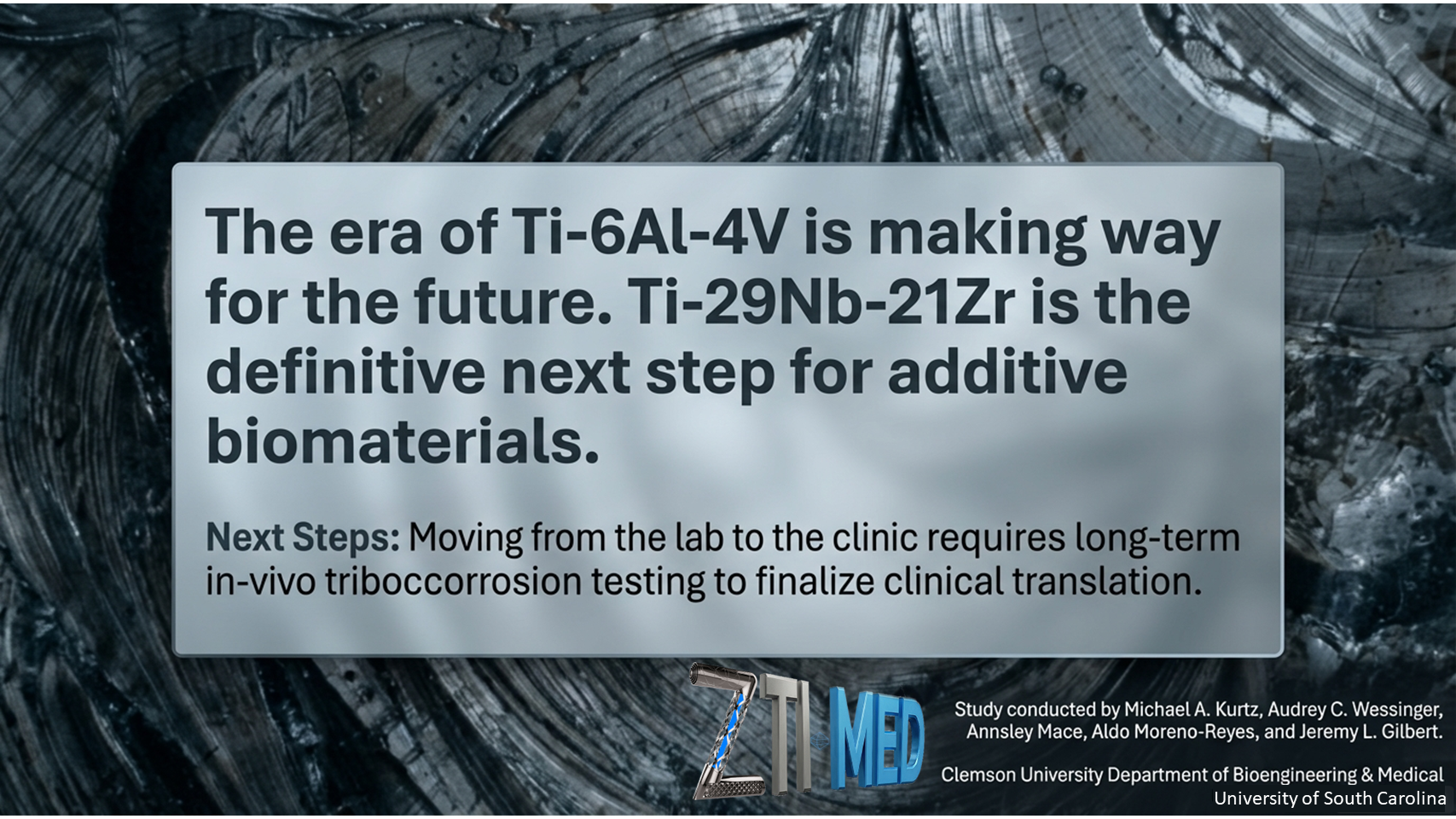
Additively manufactured Ti-29Nb-21Zr shows improved oxide polarization resistance versus Ti-6Al-4V in inflammatory simulating solution
Abstract
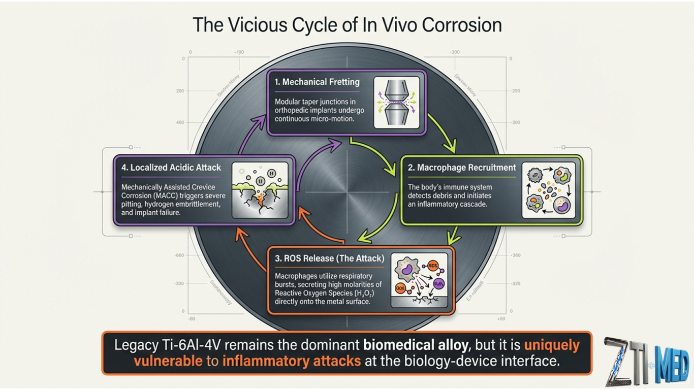
Retrieval studies in the past two decades show severe corrosion of titanium and its alloys in orthopedic implants. This damage is promoted by mechanically assisted crevice corrosion (MACC), particularly within modular titanium-titanium junctions. During MACC, titanium interfaces may be subject to negative potentials and reactive oxygen species (ROS), generated from cathodic activation and/or inflammation. Additive manufacturing (AM) may be able to produce new, corrosion-resistant titanium alloys and admixtures that are less susceptible to these adverse electrochemical events. In this study, we characterize the impedance and corrosion properties of three new AM titanium materials, including Ti-6Al-4V with added 1% nano-yttria stabilized ZrO2, admixed Ti-29Nb-21Zr, and pre-alloyed Ti-29Nb-21Zr. We aim to elucidate how these materials perform when subjected to high ROS solutions. We include conventionally and additively manufactured Ti-6Al-4V in our study as comparison groups. A 0.1 M H2O2 phosphate-buffered saline (PBS) solution, simulating inflammatory conditions, significantly increased biomaterial OCP (−0.14 V vs. Ag/AgCl) compared to PBS only (−0.38 V, p = .000). During anodic polarization, Ti-6Al-4V passive current density more than doubled from 1.28 × 10⁻⁷ to 3.81 × 10⁻⁷ A/cm2 when exposed to 0.1 M H2O2. In contrast, Ti-29Nb-21Zr passive current density remained relatively unchanged, slightly increasing from 7.49 × 10⁻⁸ in PBS to 9.31 × 10⁻⁸ in 0.1 M H2O2. Ti-29Nb-21Zr oxide polarization resistance (Rp) was not affected by 0.1 M H2O2, maintaining a high value (1.09 × 10⁶ vs. 1.89 × 10⁶ Ω cm2), while Ti-6Al-4V in 0.1 M H2O2 solution had significantly diminished Rp (4.38 × 10⁶ in PBS vs. 7.24 × 10⁴ Ω cm2 in H2O2). These results indicate that Ti-29Nb-21Zr has improved corrosion resistance in ROS containing solutions when compared with Ti-6Al-4V based biomaterials.
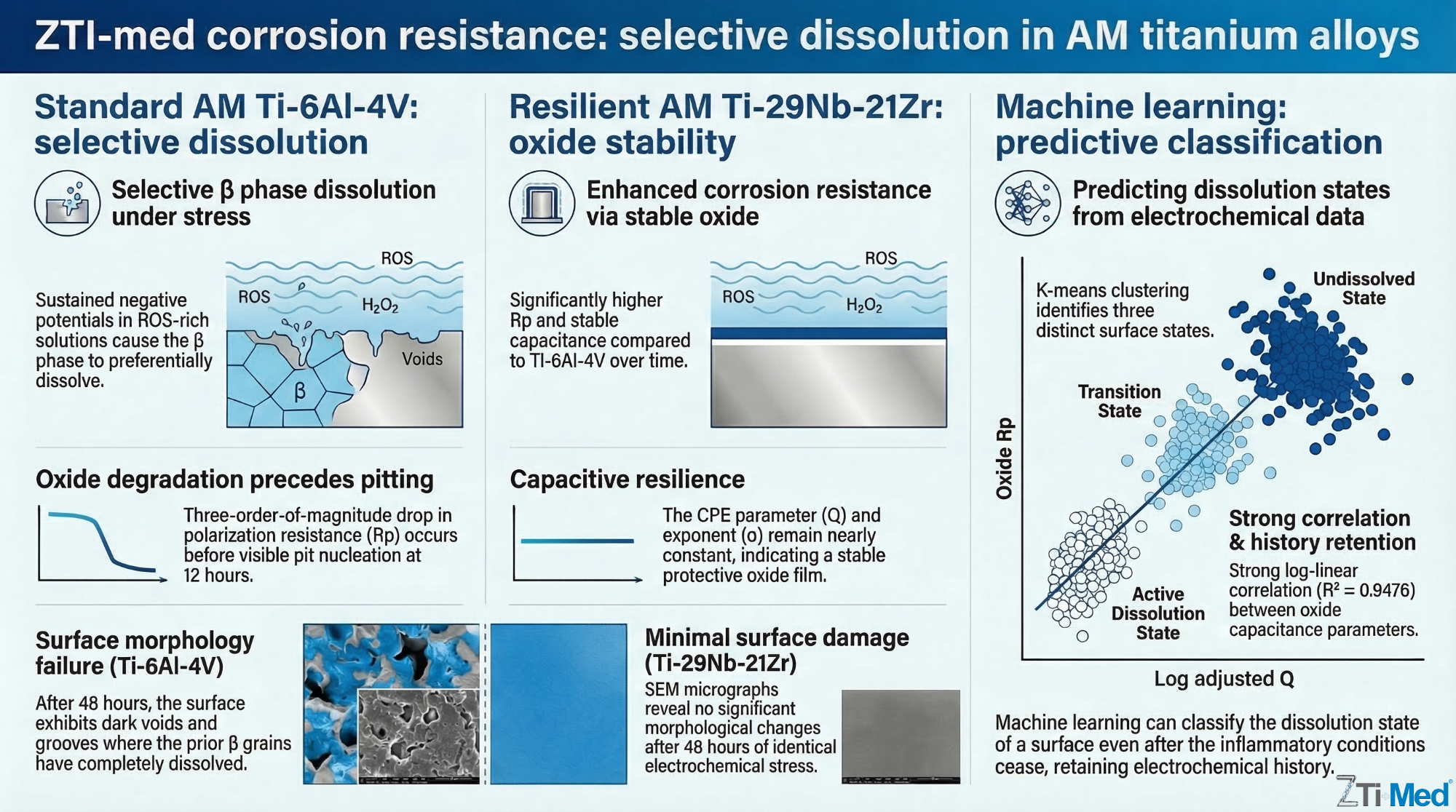
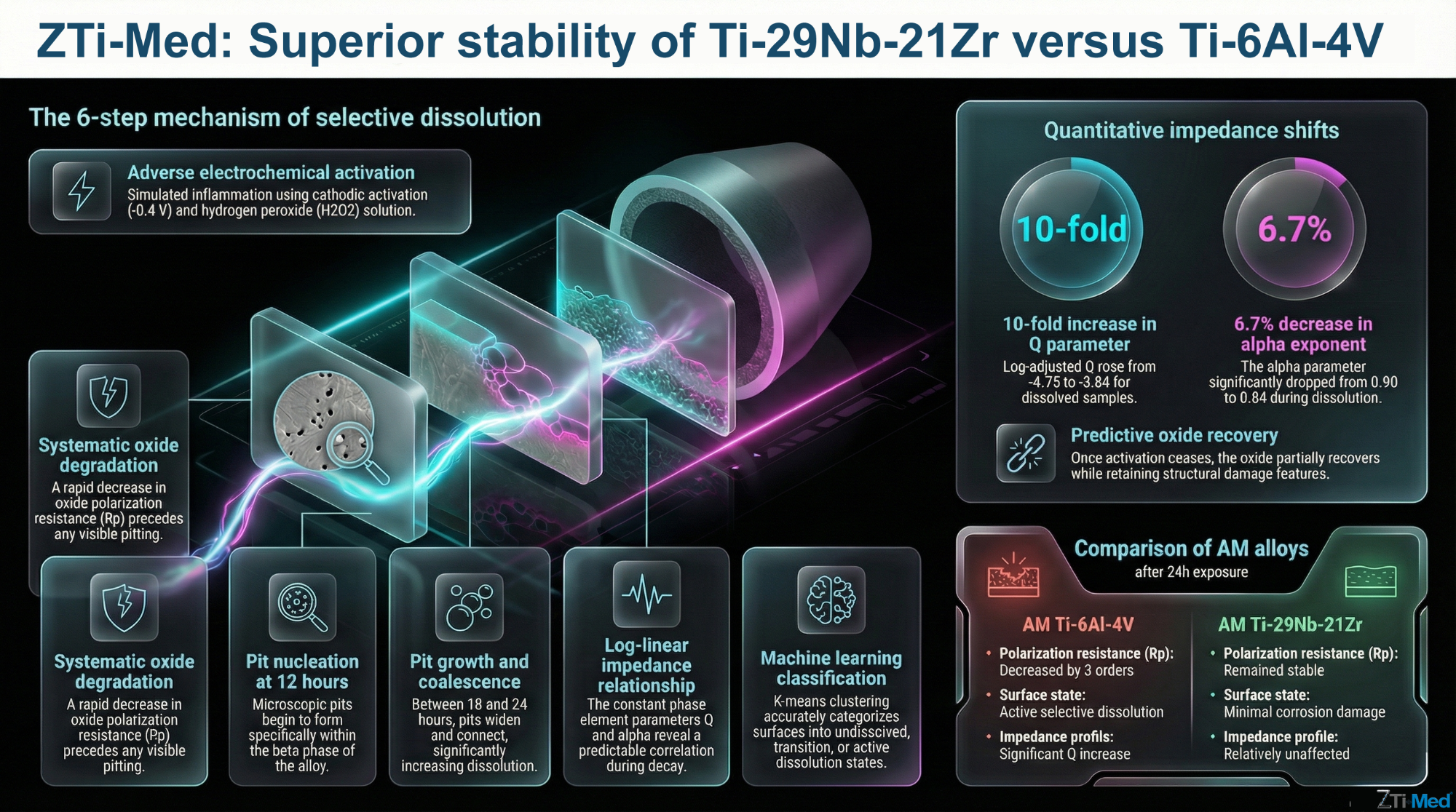
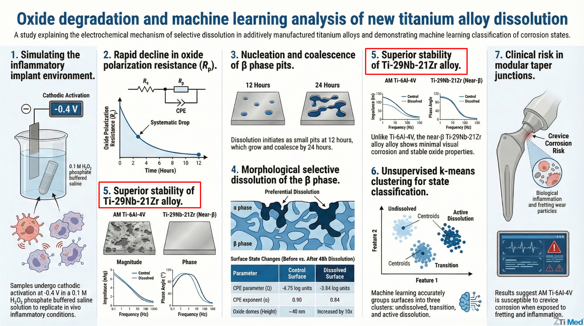
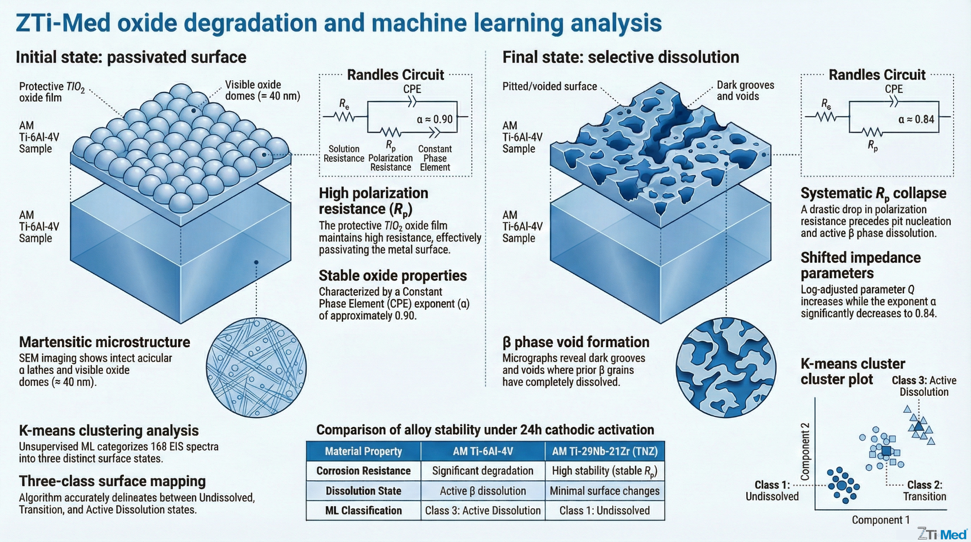
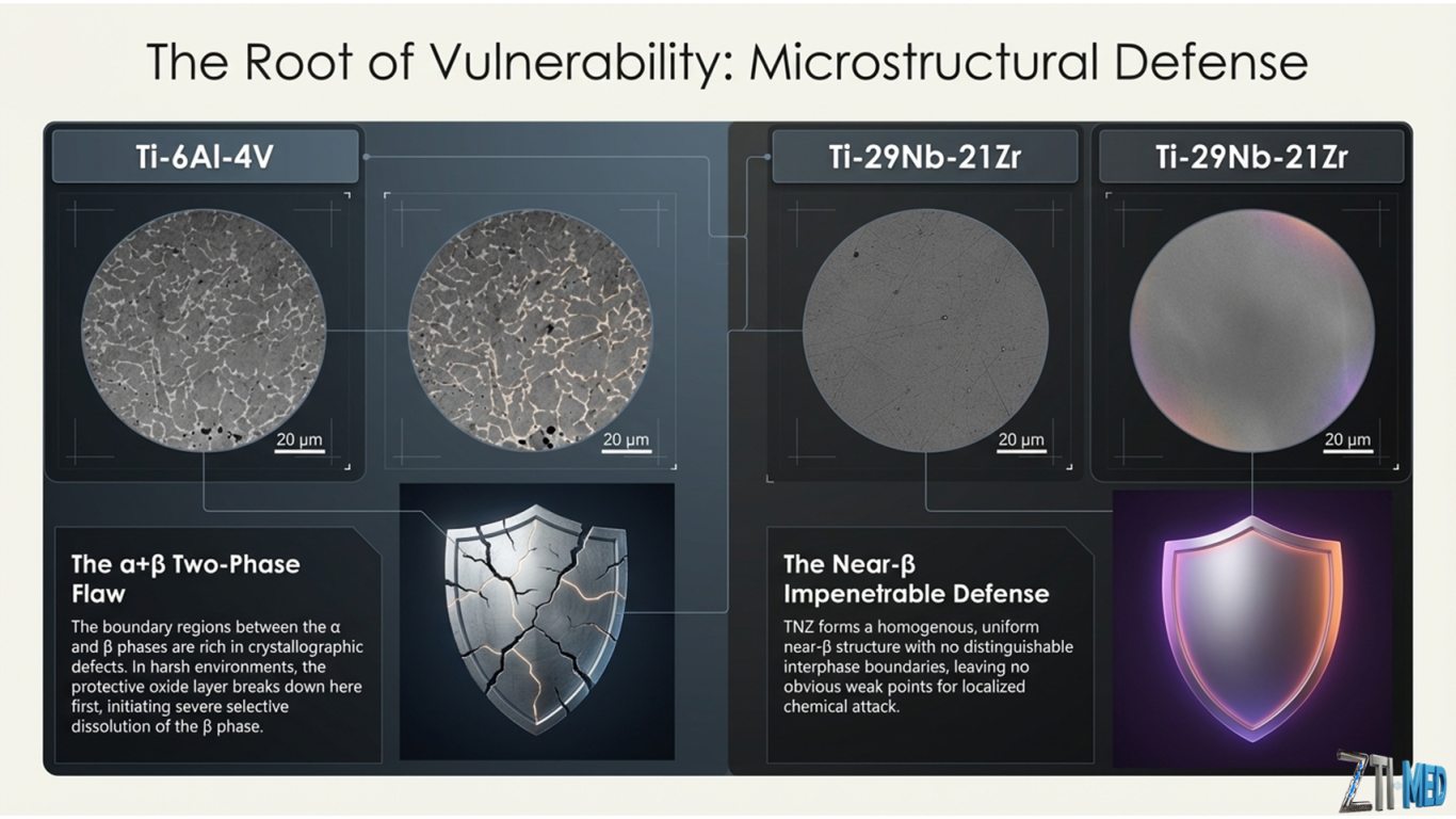
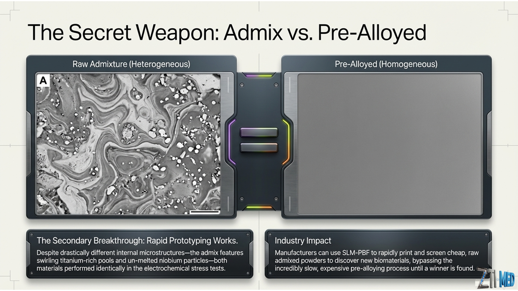
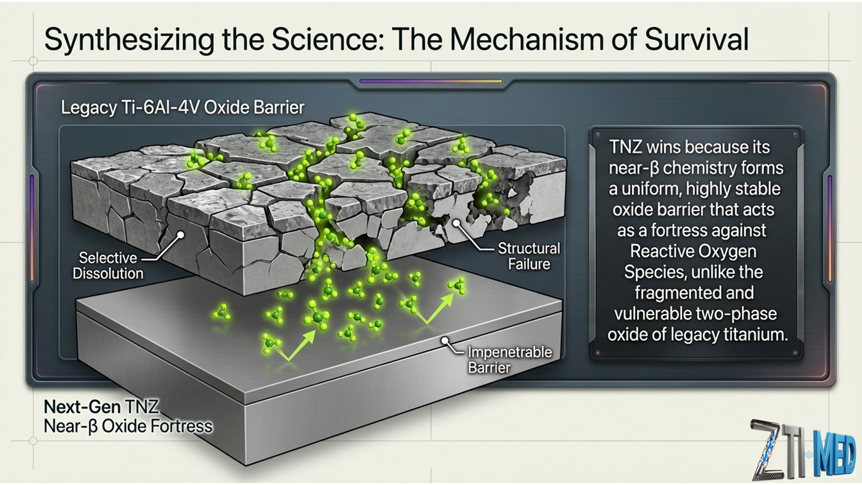
Oxide degradation precedes additively manufactured Ti-6Al-4V selective dissolution: An unsupervised machine learning correlation of impedance and dissolution compared to Ti-29Nb-21Zr
Abstract
Additively manufactured (AM) Ti-6Al-4V devices are implanted with increasing frequency. While registry data report short-term success, a gap persists in our understanding of long-term AM Ti-6Al-4V corrosion behavior. Retrieval studies document β phase selective dissolution on conventionally manufactured Ti-6Al-4V devices. Researchers reproduce this damage in vitro by combining negative potentials (cathodic activation) and inflammatory simulating solutions (H2O2-phosphate buffered saline). In this study, we investigate the effects of these adverse electrochemical conditions on AM Ti-6Al-4V impedance and selective dissolution. We hypothesize that cathodic activation and H2O2 solution will degrade the oxide, promoting corrosion. First, we characterized AM Ti-6Al-4V samples before and after a 48 h −0.4 V hold in 0.1 M H2O2/phosphate buffered saline. Next, we acquired nearfield electrochemical impedance spectroscopy (EIS) data. Finally, we captured micrographs and EIS during dissolution. Throughout, we used AM Ti-29Nb-21Zr as a comparison. After 48 h, AM Ti-6Al-4V selectively dissolved. Ti-29Nb-21Zr visually corroded less. Structural changes at the AM Ti-6Al-4V oxide interface manifested as property changes to the impedance. After dissolution, the log-adjusted constant phase element (CPE) parameter, Q, significantly increased from −4.75 to −3.84 (Scm−2(s)α) (p = .000). The CPE exponent, α, significantly decreased from .90 to .84 (p = .000). Next, we documented a systematic decrease in oxide polarization resistance before pit nucleation and growth. Last, using k-means clustering, we established a structure–property relationship between impedance and the surface's dissolution state. These results suggest that AM Ti-6Al-4V may be susceptible to in vivo crevice corrosion within modular taper junctions.

Corrosion Resistance of Additively Manufactured Titanium Alloys in Physiological and Inflammatory-Simulating Environments: Ti-6al-4v Versus Ti-29nb-21zr
Abstract
Retrieval studies in the past two decades show severe corrosion of titanium and its alloys in orthopedic implants. This damage is associated with clinical failure, resulting in a need to identify new biomaterials that are load-bearing, wear-resistant, and more corrosion-resistant. Additive manufacturing (AM) can produce new metallic admixtures and alloys that may not otherwise be producible using conventional manufacturing methods. In this study, we characterize the fundamental corrosion properties of three new AM titanium materials, including Ti-6Al-4V with added 1% nano-yttria stabilized ZrO2, admix Ti-29Nb-21Zr, and pre-alloyed Ti-29Nb-21Zr. We compare these properties to conventionally and additively manufactured Ti-6Al-4V. A 0.1 M H2O2 phosphate-buffered saline (PBS) solution, simulating inflammatory conditions, significantly increased biomaterial OCP (-0.14 V vs Ag/AgCl) compared to PBS only (-0.38 V, p=0.000). During anodic polarization, Ti-6Al-4V passive current density more than doubled from 1.28*10-7 to 3.81*10-7 A/cm2 when exposed to 0.1 M H2O2. In contrast, Ti-29Nb-21Zr passive current density remained relatively unchanged, slightly increasing from 7.49*10-8 in PBS to 9.31*10-8 in 0.1M H2O2. Ti-29Nb-21Zr oxide polarization resistance (Rp) was not affected by 0.1M H2O2, maintaining a high value (1.09 *106 vs. 1.89*106 Ωcm2), while Ti-6Al-4V in 0.1 M H2O2 solution had significantly diminished Rp (4.38*106 in PBS vs. 7.24*104 Ωcm2 in H2O2). These results indicate that Ti-29Nb-21Zr has improved corrosion resistance in inflammatory simulating environments compared to Ti-6Al-4V based biomaterials.


Notre Histoire et Notre Mission
Chez zti-med, nous avons créé ZTM14N pour répondre aux besoins critiques du secteur médical. Notre mission est de fournir des matériaux biocompatibles exempts de toxines tout en offrant un module d’élasticité adapté à l’os humain, favorisant ainsi une meilleure intégration et durabilité des implants.
Rencontrez Notre Équipe d’Experts
Notre équipe multidisciplinaire allie expertise en métallurgie, biomatériaux et technologies de fabrication additive pour développer des alliages comme ZTM14N. Nous collaborons étroitement avec des professionnels du médical pour garantir une innovation utile et fiable.